Sunlight-Driven Photothermal Effect of Composite Eggshell Membrane Coated with Graphene Oxide and Gold Nanoparticles


1
National Engineering Laboratory for Advanced Yarn and Fabric Formation and Clean Production, Wuhan Textile University, Wuhan 430073, China
2
Institute for Frontier Materials, Deakin University, Geelong/Melbourne, VIC 3216, Australia
3
Hubei Collaborative Innovation Center for Advanced Organic Chemical Materials & Ministry-of-Education Key Laboratory for the Synthesis and Application of Organic Functional Molecules & College of Chemistry and Chemical Engineering, Hubei University, Wuhan 430062, China
*
Authors to whom correspondence should be addressed.
Appl. Sci. 2019, 9(20), 4384; https://doi.org/10.3390/app9204384
Submission received: 23 September 2019
/
Revised: 14 October 2019
/
Accepted: 14 October 2019
/
Published: 17 October 2019
(This article belongs to the Special Issue Nanocomposites for the Sustainable Environment)
Abstract
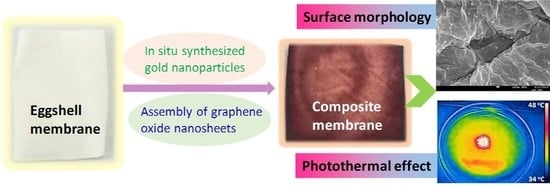
:Eggshell membrane (ESM), which consists of unique interwoven shell membrane fibers, provides a unique supporting platform for functional nanoparticles in catalysis and sensing. This work reports a novel strategy for fabricating sunlight-driven photothermal conversion composite membranes by loading graphene oxide (GO) and gold nanoparticles (AuNPs) on the three-dimension (3D) network structured eggshell membrane. Surface morphologies and chemical elements were characterized by scanning electron microscopy and X-ray photoelectron spectroscopy. High photothermal conversion under simulated sunlight irradiation, which may be caused by the synergistic effect of GO and AuNPs, was achieved by coating both GO and AuNPs onto ESM. The temperature of ESM modified with AuNPs, and then GO increased from 26.0 °C to 49.0 °C after 10 min of light irradiation. Furthermore, the nanoscaled GO and AuNPs could add benefit to the heating localization of the obtained composite membrane. It is expected this biocompatible ESM modified with GO and AuNPs would have great potential in drug release and photothermal therapy applications.
1. Introduction
Sunlight-based photothermal materials have attracted increasing attention due to the growing global demands for energy and incumbent environmental concerns. Developing effective photothermal materials is very important for various practical applications, such as seawater desalination, photothermal therapy, and drug release [1,2,3,4]. The photothermal effect is closely related to the light absorbance of materials. Plasmonic materials, including gold nanoparticles (AuNPs) and silver nanoparticles (AgNPs), show particular light absorbance at certain wavelengths. These plasmonic materials have been used as photothermal elements in many research works due to their unique localized surface plasmon resonance (LSPR) properties, which are sensitive to the particle shape, size, and metal materials. Incident light, at a certain frequency, leads to conduction electrons locally oscillating around nanoparticles. This phenomenon of excitation of surface plasmons by light irradiation is known as LSPR [5]. The plasmonic effect of AuNPs has been utilized to accelerate water evaporation and harvest sunlight energy through LSPR-based photothermal conversion [6]. Surface plasmon renders high photothermal conversion efficiency. The photo energy absorbed by plasmonic nanoparticles can be converted to the resonance energy of free electron oscillations and then transformed to lattice vibration energy [7]. This vibration energy is then transferred in the form of heat, resulting in an increase in the surrounding temperature. The small scale of the nanoparticles makes heating the area confined in the sub-micrometer area, overcoming the difficulty of heating localization from traditional methods. The local formation of a transient nanobubble around gold nanospheres in water was observed to illuminate the photothermal effect of plasmonic nanoparticles [8]. Palermo et al. investigated the LSPR induced heat generation from a monolayer one-dimensional (1D) grating of AuNPs prepared by photo-reduction in a poly (vinyl alcohol) (PVA) matrix doped with HAuCl4 onto a glass substrate, under resonant laser radiation [9]. Comprehensive theoretical modeling was developed to study the plasmonic heat production from a single layer of randomly distributed AuNPs and compared with the experimental results [10]. The influencing factors of the photothermal conversion of AuNPs solutions, including nanoparticle size, the exciting laser intensity, and the presence of guest dye molecules, were also investigated [11].
In addition to plasmonic materials, two-dimensional (2D) nanomaterials, such as graphene oxide (GO), not only exhibit great thermal conducive performance but also possess high photothermal conversion efficiency [12,13]. The photothermal features of GO have been used for water vapor generation [14] and disease therapy [13] as well as drug delivery [15]. Sunlight-induced water evaporation was realized based on the photothermal effect to GO [16]. Other functional materials, such as gold nanoparticles [17], tricalcium silicate [18], metal oxides [19], and upconverting nanomaterials [20], were combined with GO to improve the photothermal ability of GO. Plasmonic nanomaterials with tunable and localized photothermal properties enhanced the photothermal effect of GO nanosheets, achieving a highly efficient harvest and utilization of sunlight [21].
Eggshell membrane (ESM), as a low-cost waste biomass material, is a promising material for adsorption of metal ions, delivery of protein enzyme, and chemical sensors [22,23,24]. ESM locates between the egg white and eggshell, including the outer layer, inner layer, and limiting layer. Among these layers, the outer and inner layers are composed of complex intercalated nanofibers with porous three-dimensional (3D) network structures [25]. ESM with semi-permeability exhibits good air and water permeability and plays an important role in nutrition delivery for the embryo. It has been demonstrated that ESM is harmless to the human body [26]. ESM showed an inhibiting effect on bacteria, which favors the long-time storage function of the composite membrane. ESM, as a natural bio-membrane, has wide potential applications in the medical field, such as therapy for burnt skin [27,28], repair of the eardrum, and treatment of rheumatism [29,30]. It would be meaningful to fabricate composite ESM with a high photothermal effect for drug release and medical therapy.
Herein, we have developed a novel strategy for fabricating photothermal biomass membranes by combining GO nanosheets with the ESM with in situ synthesized AuNPs. ESM acted as a reducing and stabilizing agent during the preparation of AuNPs by heating. The 3D network fibrous structures of ESM provided porous supports for the GO nanosheets and AuNPs. GO nanosheets wrapped around ESM microfibers, which did not influence the in situ synthesis of AuNPs. The photothermal effects of the ESM modified with GO and AuNPs were studied by monitoring the temperature changes in the ESM surface under simulated sunlight. The synergistic photothermal effect of GO and AuNPs are discussed.
2. Materials and Methods
2.1. Materials
Tetrachloroauric (III) acid (HAuCl4·4H2O, Au content ≥47.8%) was purchased from Aladdin (Shanghai, China). GO nanosheets with a thickness of 0.8 to 1.2 nm and two-dimensional length of 0.5 to 5 μm were obtained from Nanjing Xianfeng Nano Science and Technology Ltd, China. All chemicals were analytic grade and used as received. Fresh eggs were obtained from a local supermarket. The ESM was carefully stripped from the raw egg and cut into small pieces (approximate 2.0 cm × 1.5 cm). The raw ESM was washed several times with deionized water and dried in air at ambient conditions.
2.2. Characterization
The morphologies of samples were characterized using a JEOL JSM-7800F field-emission scanning electron microscope (SEM) (JEOL Ltd., Tokyo, Japan). Thermogravimetric analysis (TGA) was carried out on a Perkin Elmer’s Diamond TG/DTA system (PerkinElmer Inc., Waltham, Massachusetts, USA), at a heating rate of 10 °C min−1 in a nitrogen atmosphere. X-ray photoelectron spectra (XPS) were observed by a Thermo Fisher Scientific 250 Xi system (Thermo Fisher Scientific, Waltham, Massachusetts, USA). UV-Vis diffuse reflectance spectra of samples were recorded using a Shimadzu SolidSpec-3700/3700DUV UV-VIS-NIR Spectrophotometer (Shimadzu Corp., Tokyo, Japan). The Renishaw INVIA Raman microscope system (Renishaw plc, Wotton-under-Edge, Gloucestershire, UK) with a 633 nm He-Ne laser excitation source (17 mW) was used for Raman analysis. A xenon lamp (λ = 350–780 nm) (CEL-HXUV 300, Beijing Education Au-light Co., Ltd., Beijing, China) was used to simulate the sunlight source for photothermal tests.
2.3. In-Situ Synthesis of AuNPs on ESM
The dry ESM was immersed in deionized water for 10 min. Each ESM sample (approximate 2.0 cm × 1.5 cm) was socked in 30 mL of HAuCl4 aqueous solution (1.5 × 10⁻⁴ M). The pH value of the reaction solution was measured to be around 6. After being shaken for 5 min at room temperature, the HAuCl4 solution with ESM was shaken for 2 h in water batch at 95 ℃. The purple–red ESM was obtained after heat treatment, denoted as AuNP/ESM.
2.4. Assembly of GO on ESM
The combination of GO and ESM was achieved by the dip-coating method. ESM was immersed in GO aqueous solution with 0.3 mg mL−1 at a liquid ratio of 1000:1. The GO solution containing ESM was shaken for 30 min at room temperature. Then the ESM was taken out and dried under ambient condition. The adsorption of GO onto ESM was repeated in triplicate. The obtained GO coated ESM was denoted as GO/ESM.
Composite films, including GO/AuNP/ESM and AuNP/GO/ESM, were fabricated by a complex assembly of GO and in situ synthesis of AuNPs on ESM. The AuNP treated ESM (AuNP/ESM) was assembled by GO and formed GO/AuNP/ESM. The GO coated ESM (GO/ESM) was modified in the HAuCl4 solution by heat treatment, and then AuNP/GO/ESM was obtained.
2.5. Photothermal Measurement of ESM
The photothermal performance of the samples was tested under simulated sunlight from the xenon lamp. The distance between samples and the xenon lamp was 20 cm. The temperatures of the ESM samples during light irradiation were recorded by a FLIR C2 compact thermal imaging camera (FLIR Systems, Inc., Wilsonville, Oregon, USA) at a time interval of 10 min.
3. Results and Discussion
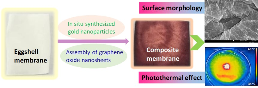
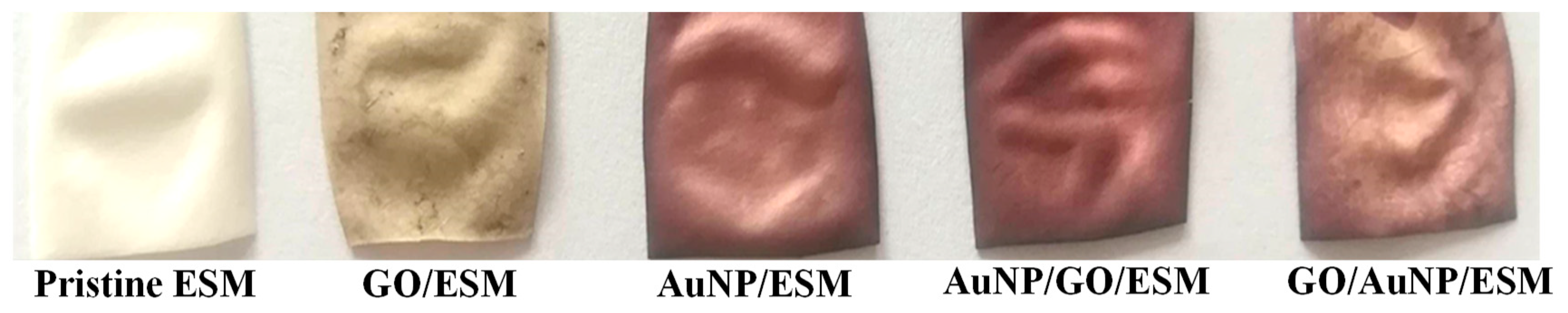
The color of ESM changed from white to brown–yellow after GO nanosheets were coated on the surface of ESM (GO/ESM in Figure 1), indicating that GO nanosheets were combined effectively with ESM. The color of ESM changed from white to wine red after the in situ synthesis of gold nanoparticles (AuNP/ESM in Figure 1). The ESM sample became black–purple when the AuNPs were in situ-synthesized onto the GO-modified ESM (Au/GO/ESM in Figure 1). The AuNP-treated ESM was further coated with GO, which showed a dark color (GO/AuNP/ESM in Figure 1). It should be noted that the color of the GO/ESM became darker during the in situ heating synthesis of AuNPs, implying that GO may be reduced by heat in the presence of ESM, which is similar to the GO reduction on the cotton surface at high temperature [31]. To investigate the optical properties of the ESM samples, UV-Vis diffusion reflectance absorbance spectra were measured (Figure 2). The pristine ESM displayed absorption in the UV region due to its protein composition. The GO/ESM showed higher absorption in the whole wavelength range compared to the pristine ESM because of the coating of GO nanosheets. The curve of AuNP/ESM presented an absorption band around 540 nm, which is assigned to the LSPR mode of AuNPs on the ESM surface [32]. A distinct absorption band around 540 nm was observed in the UV-Vis diffusion reflectance absorbance spectra of AuNP/GO/ESM and GO/AuNP/ESM, indicating that the GO coating did not obviously change the LSPR properties of AuNPs. The complex of GO and AuNPs enhanced the UV-Vis absorption of the ESM samples. The spectral data indicates that GO nanosheets were coated on ESM surface, and AuNPs were in situ-synthesized on ESM after heat treatment.
Surface morphologies of the ESM samples were observed by SEM. Pristine ESM displays 3D nonwoven network with porous structures (Figure 3a). The average diameter of the fibers in ESM was measured to be 2.00 μm (Figure 3b). After treatment with GO, numerous wrinkle nanosheets were found wrapped around ESM fibers, which indicates that GO has been effectively coated onto the networks of ESM (Figure 3c,d). No GO nanosheets were observed in the pores in the 3D structures of ESM, implying direct interaction between ESM fibers and GO nanosheets (Figure 3c). The oxygen-containing groups on the GO nanosheets interacted with the amino and carboxyl groups from amino acids on ESM, which leads to the effective coating of GO nanosheets around the fibrous structures of ESM. As can be seen from the SEM images of AuNP/ESM (Figure 3e,f), a dense coating of nanoparticles was observed on the surface of the ESM network fibers. The average size of the nanoparticles was measured to be 21.15 nm (Figure 3f). The SEM images clearly demonstrate that AuNPs were synthesized onto the ESM surface. The diverse amino acids existing in ESM could possess a reducing effect [33,34], which may result in the reduction of Au ions and then the formation of AuNPs on the ESM by heat treatment [35]. A large number of AuNPs was also observed in the SEM images of AuNP/GO/ESM (Figure 3g) and GO/AuNP/ESM (Figure 3h). The presence of GO nanosheets did not affect the in situ synthesis of AuNPs. In the composite films, wrinkled GO nanosheets and spherical AuNPs were observed obviously on the fibrous structures of ESM. The average sizes of AuNPs on AuNP/GO/ESM and GO/AuNP/ESM were 26.76 and 22.47 nm, respectively. There was no obvious size change in the AuNPs after the assembly of GO on the AuNP/ESM. Nevertheless, this is compared with an increased AuNPs size when Au was in situ-synthesized onto GO/ESM. GO may promote the synthesis of AuNPs due to the active groups on the surface of GO nanosheets.
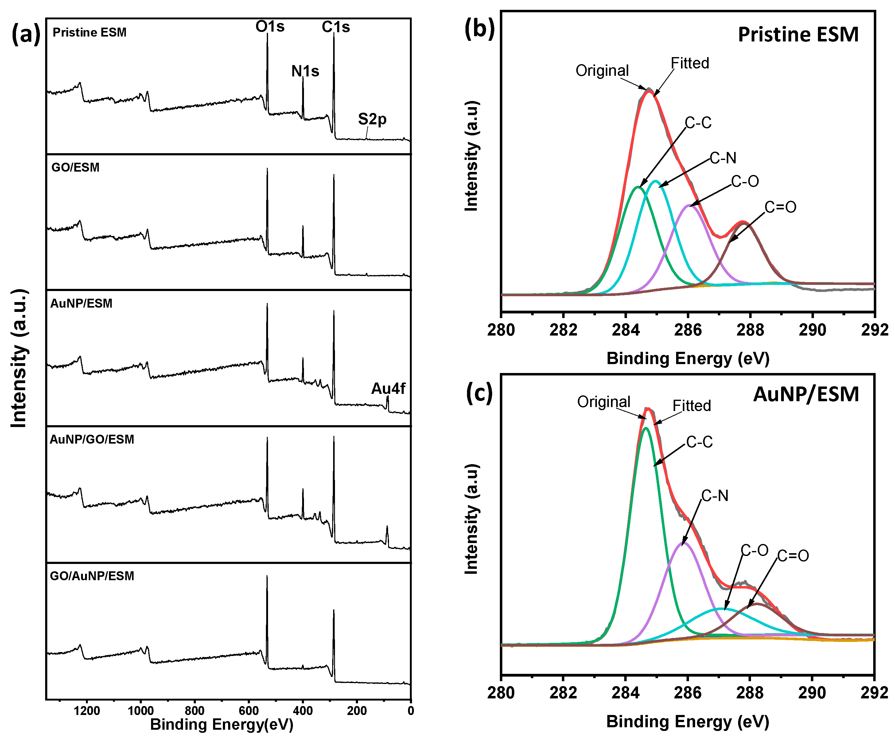
The surface chemical elements of different ESM samples were inspected by XPS. XPS survey spectra of the ESM samples displayed peaks at 284.8, 399.68, 163.62, and 531.28 eV, corresponding to C1s, N1s, S2p, and O1s, respectively. These are the elements of protein-based materials (Figure 4a). Furthermore, there is a peak at 83.86 eV assigned to Au4f, appearing in the XPS survey curves of AuNP/GO/ESM and AuNP/ESM, implying that AuNPs were prepared on the surface of ESM samples. After peak fitting, the C1s XPS band of pristine ESM can be fitted into four peaks at 284.39, 284.94, 286.05, and 287.79 eV (Figure 4a), which are ascribed to C–C, C–N, C–O, and C=O, respectively [36,37]. The C1s XPS band of AuNP/ESM also shows four fitting peaks at 284.65, 285.86, 287.08, and 288.22 eV (Figure 4b). The synthesis of AuNPs led to a slight increase in the binding energy of carbon bonds. It is clearly observed that the proportion of C–C increased, and the proportion of C–O and C=O decreased in AuNP/ESM compared with pristine ESM (Figure 4c), which could be attributed to aldehydelation of ESM. The AuNPs may be formed through the reduction of Au ions by aldehydelation of ESM [38].
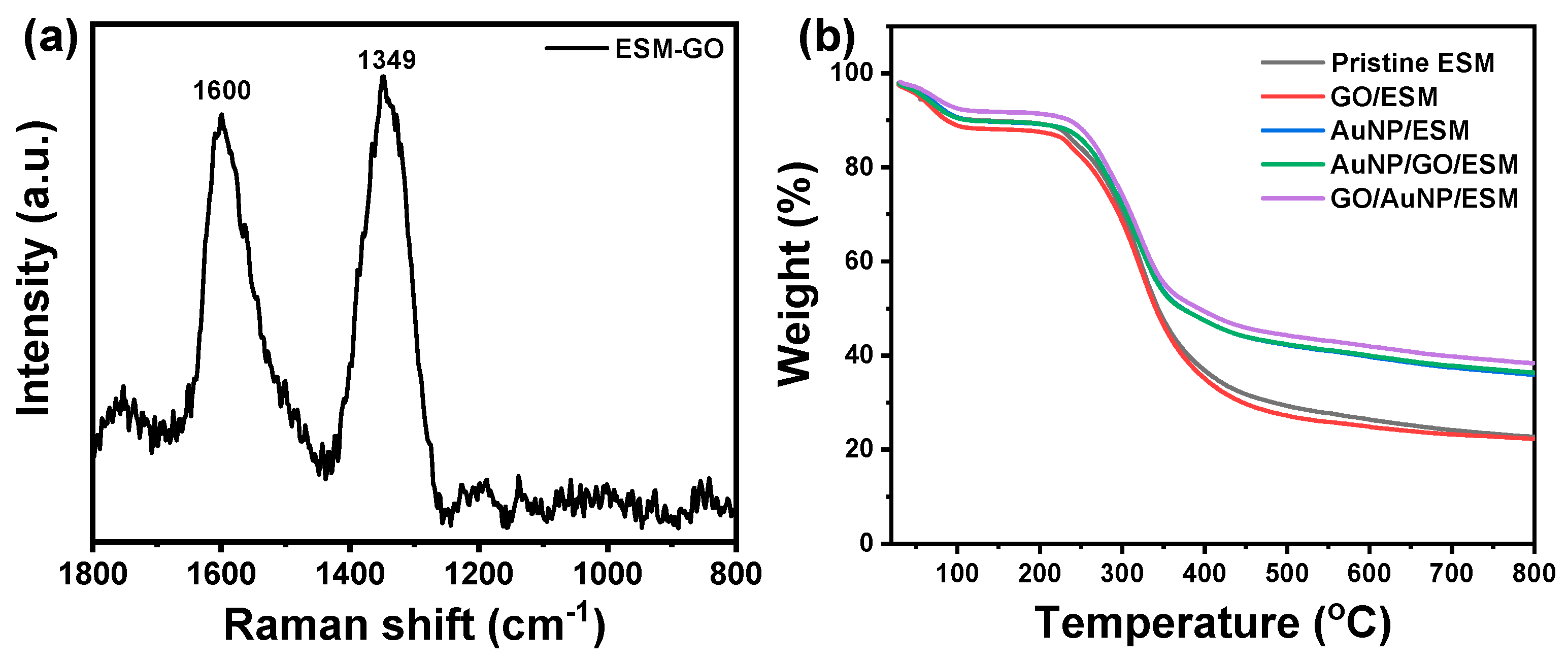
The coating of GO on the surface of ESM was further confirmed by Raman scattering spectroscopy. As shown in Figure 5a, two Raman bands were observed at 1349 and 1600 cm−1, which corresponded to the in-plane bond stretching motion of C sp2 atoms (G band) and the breathing modes of benzenoid rings (D band) of GO [39]. The result further demonstrated the successful coating of GO nanosheets on ESM. The composition and thermal stability of ESM samples were tested by TGA. The weight loss of the samples below 100 °C is due to the evaporation of the moisture inside samples (Figure 5b). A major weight loss was observed for all the samples and was found from 240 °C to 400 °C, which was attributed to the decomposition of ESM. There was only a slight weight loss with a further increase in the temperature, which indicates the thermal decomposition of the ESM samples was nearly complete at around 400 °C. The residual percentages for pristine ESM, GO/ESM, AuNP/ESM, AuNP/GO/ESM, and GO/AuNP/ESM were 22.73%, 22.31%, 35.92%, 36.39%, and 38.37%, respectively. The ESM samples with the coating of both the AuNPs and GO nanosheets showed higher residual percentages compared to other samples, implying that more nanoparticles remained in the residuals.
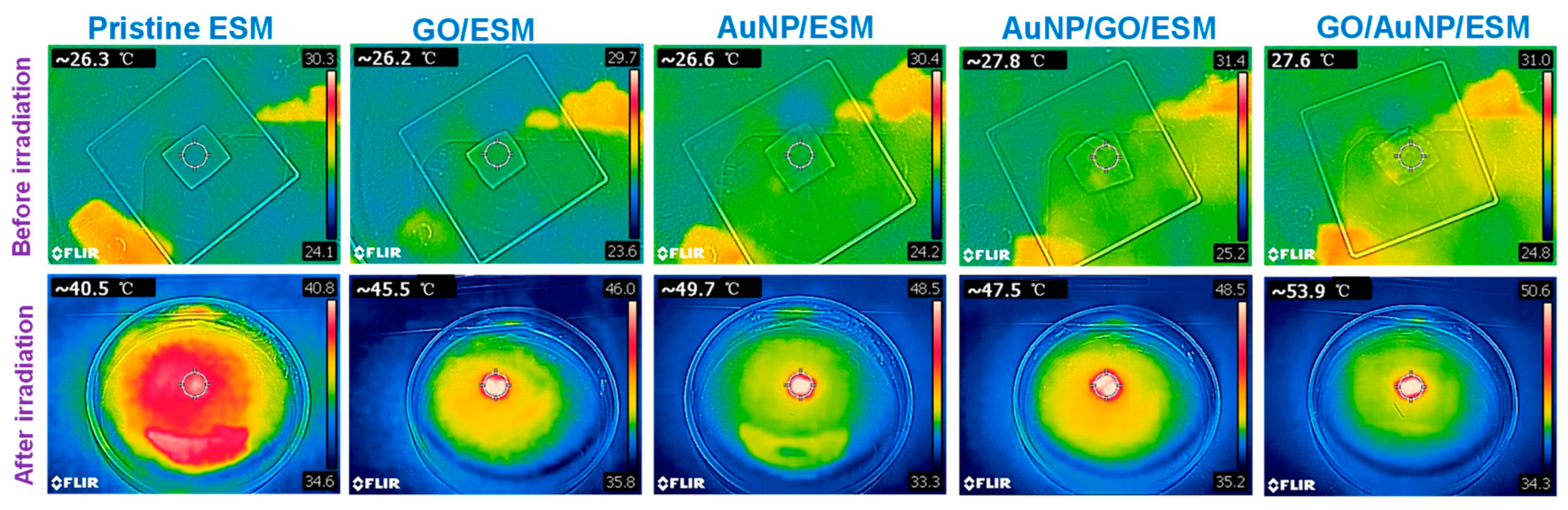
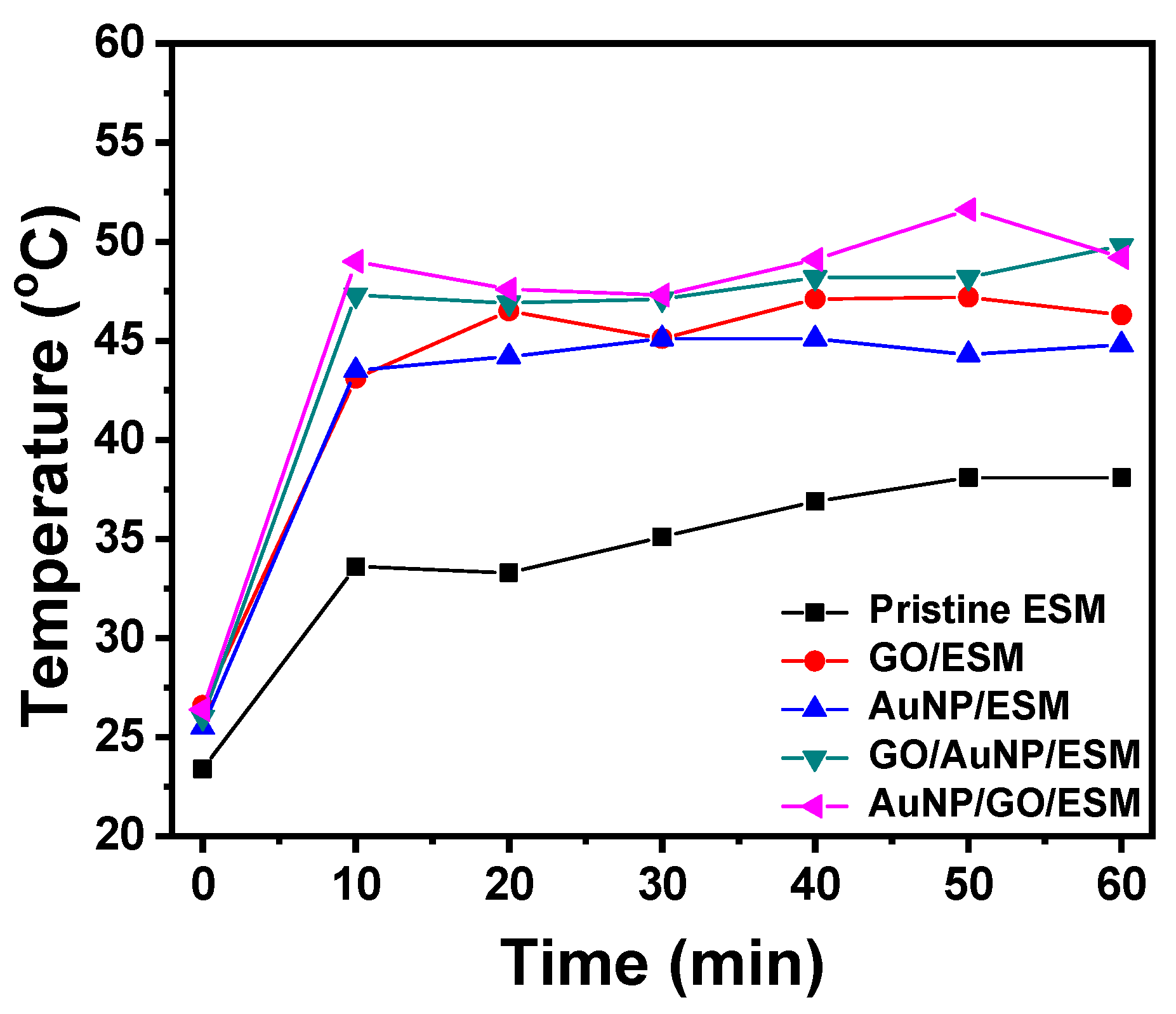
The photothermal effect of the ESM samples was investigated by being subjected to simulated sunlight irradiation. Figure 6 displays the infrared thermal images of the ESM samples before and after light irradiation for 10 min under the xenon lamp. The surface temperatures of the ESM samples increased greatly under the light irradiation. The temperature of the pristine ESM increased from 23.4 to 33.6 °C after light irradiation of 10 min. This is compared with a much higher temperature achieved by GO/ESM, AuNP/ESM, AuNP/GO/ESM, and GO/AuNP/ESM under the same conditions, which was 43.1, 43.5, 47.3, and 49.0 °C, respectively. The thermal images show that the heat distribution of the pristine ESM was over the whole sample as well as the other irradiated area. With photothermal conversion materials, the GO and AuNPs coated ESM showed a concentrated small heat distribution area in the middle of the samples, which suggests that the ESM coated with GO and AuNPs could be used for localized sunlight driven heating. The temperatures of ESM samples varied slightly as the irradiation time was further prolonged (Figure 7), implying that the photothermal conversion reached equilibrium within 10 min. The rapid photothermal conversion of ESM samples facilitates the applications of GO and AuNPs coated ESM. ESM coated with both GO and AuNPs exhibited higher temperatures (51.6 °C) than the ESM with GO or AuNPs only, which reveals that the synergistic effect of GO and AuNPs increased the photothermal efficiency of ESM. The LSPR band of AuNPs located around 540 nm, which was in favor of the sunlight absorption in the visible region.
4. Conclusions
In summary, photothermal biofilm was successfully fabricated by coating eggshell membrane (ESM) with graphene oxide (GO) nanosheets and gold nanoparticles (AuNPs) together. AuNPs were formed in situ on ESM that acted as a reducing and stabilizing agent. Moreover, ESM with 3D network structures played a vital supporting role for the prepared AuNPs. The presence of GO on ESM did not influence the in situ-synthesis of AuNPs. The combining effects of GO and AuNPs endowed ESM with a highly effective photothermal property. Among the ESM samples, AuNP/GO/ESM exhibited the best photothermal conversion ability, reaching 49.0 °C after being irradiated for 10 min under the simulated sunlight. Because of the compatibility of ESM and the photothermal effect of GO and AuNPs, this as-prepared photothermal composite biomass membrane could be applied for photo-driven medicine release and photothermal disease therapy.
Author Contributions
Conceptualization, B.T. and J.W.; methodology, L.W. and J.Z.; formal analysis, L.W. and H.Z.; investigation L.W., J.Z., and H.Z.; funding acquisition, B.T.; writing—original draft preparation, L.W. B.T., and J.W.; writing—review and editing, W.C. and J.W.; supervision, B.T.
Funding
This research was funded by the Hubei Provincial Natural Science Foundation of China (2018CFB523).
Conflicts of Interest
The authors declare no conflict of interest.
References
- Zhou, L.; Tan, Y.L.; Wang, J.Y.; Xu, W.C.; Yuan, Y.; Cai, W.S.; Zhu, S.N.; Zhu, J. 3D self-assembly of aluminium nanoparticles for plasmon-enhanced solar desalination. Nat. Photonics 2016, 10, 393–398. [Google Scholar] [CrossRef]
- Austin, L.A.; Mackey, M.A.; Dreaden, E.C.; El-Sayed, M.A. The optical, photothermal, and facile surface chemical properties of gold and silver nanoparticles in biodiagnostics, therapy, and drug delivery. Arch. Toxicol. 2014, 88, 1391–1417. [Google Scholar] [CrossRef] [PubMed] [Green Version]
- Yang, J.; Shen, D.; Zhou, L.; Li, W.; Li, X.; Yao, C.; Wang, R.; El-Toni, A.M.; Zhang, F.; Zhao, D. Spatially confined fabrication of core-shell gold nanocages@mesoporous silica for near-infrared controlled photothermal drug release. Chem. Mater. 2013, 25, 3030–3037. [Google Scholar] [CrossRef]
- Ricciardi, L.; Sancey, L.; Palermo, G.; Termine, R.; De Luca, A.; Szerb, E.I.; Aiello, I.; Ghedini, M.; Strangi, G.; La Deda, M. Plasmon-mediated cancer phototherapy: The combined effect of thermal and photodynamic processes. Nanoscale 2017, 9, 19279–19289. [Google Scholar] [CrossRef]
- Kelly, K.L.; Coronado, E.; Zhao, L.L.; Schatz, G.C. The optical properties of metal nanoparticles: The influence of size, shape, and dielectric environment. J. Phys. Chem. B 2003, 107, 668–677. [Google Scholar] [CrossRef]
- Chang, C.; Yang, C.; Liu, Y.M.; Tao, P.; Song, C.Y.; Shang, W.; Wu, J.B.; Deng, T. Efficient solar-thermal energy harvest driven by interfacial plasmonic heating-assisted evaporation. ACS Appl. Mater. Interfaces 2016, 8, 23412–23418. [Google Scholar] [CrossRef]
- Smith, J.G.; Faucheaux, J.A.; Jain, P.K. Plasmon resonances for solar energy harvesting: A mechanistic outlook. Nano Today 2015, 10, 67–80. [Google Scholar] [CrossRef]
- Metwally, K.; Mensah, S.; Baffou, G. Fluence threshold for photothermal bubble generation using plasmonic nanoparticles. J. Phys. Chem. C 2015, 119, 28586–28596. [Google Scholar] [CrossRef]
- Palermo, G.; Ritacco, T.; Aceti, D.M.; Pezzi, L.; Giocondo, M.; De Luca, A. Photo-thermal effects in 1D gratings of gold nanoparticles. Crystals 2017, 7, 14. [Google Scholar] [CrossRef]
- Pezzi, L.; Palermo, G.; Veltri, A.; Cataldi, U.; Bürgi, T.; Ritacco, T.; Giocondo, M.; Umeton, C.; De Luca, A. Photo-thermal study of a layer of randomly distributed gold nanoparticles: From nano-localization to macro-scale effects. J. Phys. D Appl. Phys. 2017, 50, 435302. [Google Scholar] [CrossRef]
- Palermo, G.; Pagnotto, D.; Ricciardi, L.; Pezzi, L.; La Deda, M.; De Luca, A. Thermoplasmonic effects in gain-assisted nanoparticle solutions. J. Phys. Chem. C 2017, 121, 24185–24191. [Google Scholar] [CrossRef]
- Tian, B.; Wang, C.; Zhang, S.; Feng, L.Z.; Liu, Z. Photothermally enhanced photodynamic therapy delivered by nano-graphene oxide. ACS Nano 2011, 5, 7000–7009. [Google Scholar] [CrossRef] [PubMed]
- Li, M.; Yang, X.J.; Ren, J.S.; Qu, K.G.; Qu, X.G. Using graphene oxide high near-infrared absorbance for photothermal treatment of alzheimer’s disease. Adv. Mater. 2012, 24, 1722–1728. [Google Scholar] [CrossRef] [PubMed]
- Fang, W.; Zhao, L.; Chen, H.; He, X.; Li, W.X.; Du, X.; Sun, Z.M.; Zhang, T.; Shen, Y. Graphene oxide foam fabricated with surfactant foaming method for efficient solar vapor generation. J. Mater. Sci. 2019, 54, 12782–12793. [Google Scholar] [CrossRef]
- Xu, Y.R.; Hu, X.L.; Guan, P.; Du, C.B.; Tian, Y.; Ding, S.C.; Li, Z.L.; Yan, C.R. A novel controllable molecularly imprinted drug delivery system based on the photothermal effect of graphene oxide quantum dots. J. Mater. Sci. 2019, 54, 9124–9139. [Google Scholar] [CrossRef]
- Li, X.; Xu, W.; Tang, M.; Zhou, L.; Zhu, B.; Zhu, S.; Zhu, J. Graphene oxide-based efficient and scalable solar desalination under one sun with a confined 2D water path. Proc. Natl. Acad. Sci. USA 2016, 113, 13953–13958. [Google Scholar] [CrossRef] [PubMed] [Green Version]
- Luo, J.W.; Deng, W.J.; Yang, F.; Wu, Z.Q.; Huang, M.T.; Gu, M.Y. Gold nanoparticles decorated graphene oxide/nanocellulose paper for NIR laser-induced photothermal ablation of pathogenic bacteria. Carbohydr. Polym. 2018, 198, 206–214. [Google Scholar] [CrossRef]
- Xu, C.; Ma, B.; Peng, J.L.; Gao, L.; Xu, Y.H.; Huan, Z.G.; Chang, J. Tricalcium silicate/graphene oxide bone cement with photothermal properties for tumor ablation. J. Mat. Chem. B 2019, 7, 2808–2818. [Google Scholar] [CrossRef]
- Cao, Y.; Hassan, M.; Cheng, Y.; Chen, Z.R.; Wang, M.; Zhang, X.Z.; Haider, Z.S.; Zhao, G. Multifunctional Photo-and Magnetoresponsive Graphene Oxide-Fe3O4 Nanocomposite-alginate hydrogel platform for ice recrystallization inhibition. ACS Appl. Mater. Interfaces 2019, 11, 12379–12388. [Google Scholar] [CrossRef] [PubMed]
- Li, Z.L.; Johnson, O.; Huang, J.; Feng, T.; Yang, C.Q.; Liu, Z.X.; Chen, W. Enhancing the photothermal conversion efficiency of graphene oxide by doping with NaYF4: Yb, Er upconverting luminescent nanocomposites. Mater. Res. Bull. 2018, 106, 365–370. [Google Scholar] [CrossRef]
- Wang, M.M.; Zhang, J.; Wang, P.; Li, C.P.; Xu, X.L.; Jin, Y.D. Bifunctional plasmonic colloidosome/graphene oxide-based floating membranes for recyclable high-efficiency solar-driven clean water generation. Nano Res. 2018, 11, 3854–3863. [Google Scholar] [CrossRef]
- Mittal, A.; Teotia, M.; Soni, R.K.; Mittal, J. Applications of egg shell and egg shell membrane as adsorbents: A review. J. Mol. Liq. 2016, 223, 376–387. [Google Scholar] [CrossRef]
- Daraei, H.; Mittal, A.; Mittal, J.; Kamali, H. Optimization of Cr (VI) removal onto biosorbent eggshell membrane: Experimental & theoretical approaches. Desalin. Water Treat. 2014, 52, 1307–1315. [Google Scholar]
- Chen, W.; Li, B.; Xu, C.; Wang, L. Chemiluminescence flow biosensor for hydrogen peroxide using DNAzyme immobilized on eggshell membrane as a thermally stable biocatalyst. Biosens. Bioelectron. 2009, 24, 2534–2540. [Google Scholar] [CrossRef] [PubMed]
- Messens, W.; Grijspeerdt, K.; Herman, L. Eggshell characteristics and penetration by Salmonella enterica serovar Enteritidis through the production period of a layer flock. Br. Poult. Sci. 2005, 46, 694–700. [Google Scholar] [CrossRef]
- Kaweewong, K.; Garnjanagoonchorn, W.; Jirapakkul, W.; Roytrakul, S. Solubilization and identification of hen eggshell membrane proteins during different times of chicken embryo development using the proteomic approach. Protein J. 2013, 32, 297–308. [Google Scholar] [CrossRef]
- Jun, H.J.; Oh, K.-H.; Yoo, J.; Han, W.-G.; Chang, J.; Jung, H.H.; Choi, J. A new patch material for tympanic membrane perforation by trauma: The membrane of a hen egg shell. Acta Otolaryngol. (Stockh.) 2014, 134, 250–254. [Google Scholar] [CrossRef]
- Ahmed, T.A.E.; Suso, H.-P.; Maqbool, A.; Hincke, M.T. Processed eggshell membrane powder: Bioinspiration for an innovative wound healing product. Mater. Sci. Eng. C-Mater. Biol. Appl. 2019, 95, 192–203. [Google Scholar] [CrossRef]
- Ruff, K.J.; Winkler, A.; Jackson, R.W.; DeVore, D.P.; Ritz, B.W. Eggshell membrane in the treatment of pain and stiffness from osteoarthritis of the knee: A randomized, multicenter, double-blind, placebo-controlled clinical study. Clin. Rheumatol. 2009, 28, 907–914. [Google Scholar] [CrossRef]
- Ruff, K.J.; DeVore, D.P.; Leu, M.D.; Robinson, M.A. Eggshell membrane: A possible new natural therapeutic for joint and connective tissue disorders. Results from two open-label human clinical studies. Clin. Interv. Aging 2009, 4, 235–240. [Google Scholar] [CrossRef] [Green Version]
- Cai, G.M.; Xu, Z.L.; Yang, M.Y.; Tang, B.; Wang, X.G. Functionalization of cotton fabrics through thermal reduction of graphene oxide. Appl. Surf. Sci. 2017, 393, 441–448. [Google Scholar] [CrossRef]
- Dreaden, E.C.; Alkilany, A.M.; Huang, X.; Murphy, C.J.; El-Sayed, M.A. The golden age: Gold nanoparticles for biomedicine. Chem. Soc. Rev. 2012, 41, 2740–2779. [Google Scholar] [CrossRef] [PubMed]
- Nakano, T.; Ikawa, N.; Ozimek, L. Chemical composition of chicken eggshell and shell membranes. Poult. Sci. 2003, 82, 510–514. [Google Scholar] [CrossRef] [PubMed]
- Yi, F.; Guo, Z.-X.; Zhang, L.-X.; Yu, J.; Li, Q. Soluble eggshell membrane protein: Preparation, characterization and biocompatibility. Biomaterials 2004, 25, 4591–4599. [Google Scholar] [CrossRef] [PubMed]
- Tang, B.; Sun, L.; Kaur, J.; Yu, Y.; Wang, X.G. In-situ synthesis of gold nanoparticles for multifunctionalization of silk fabrics. Dyes Pigm. 2014, 103, 183–190. [Google Scholar] [CrossRef]
- Wang, Q.; Jiang, Z.; Wang, Y.; Chen, D.; Yang, D. Photocatalytic properties of porous C-doped TiO2 and Ag/C-doped TiO2 nanomaterials by eggshell membrane templating. J. Nanopart. Res. 2009, 11, 375–384. [Google Scholar] [CrossRef]
- Wang, Q.; Liu, X.; Zhang, L.; Lv, Y. Microwave-assisted synthesis of carbon nanodots through an eggshell membrane and their fluorescent application. Analyst 2012, 137, 5392–5397. [Google Scholar] [CrossRef]
- Zheng, B.; Qian, L.; Yuan, H.; Xiao, D.; Yang, X.; Paau, M.C.; Choi, M.M.F. Preparation of gold nanoparticles on eggshell membrane and their biosensing application. Talanta 2010, 82, 177–183. [Google Scholar] [CrossRef]
- Balakumar, V.; Prakash, P. A facile in situ synthesis of highly active and reusable ternary Ag-PPy-GO nanocomposite for catalytic oxidation of hydroquinone in aqueous solution. J. Catal. 2016, 344, 795–805. [Google Scholar] [CrossRef]
Figure 1.
Optical image of eggshell membrane (ESM) samples.

Figure 2.
UV-Vis diffusion reflectance absorbance spectra of the treated ESM samples.

Figure 3.
SEM images of different ESM samples: (a,b) pristine ESM, (c,d) GO/ESM, (e,f) AuNP/ESM, (g) AuNP/GO/ESM, and (h) GO/AuNP/ESM.
Figure 3.
SEM images of different ESM samples: (a,b) pristine ESM, (c,d) GO/ESM, (e,f) AuNP/ESM, (g) AuNP/GO/ESM, and (h) GO/AuNP/ESM.

Figure 4.
XPS spectra of ESM samples: (a) survey curves, and C1s XPS curve of (b) pristine ESM and (c) AuNP/ESM.
Figure 4.
XPS spectra of ESM samples: (a) survey curves, and C1s XPS curve of (b) pristine ESM and (c) AuNP/ESM.

Figure 5.
(a) Raman scattering spectrum of GO/ESM. (b) TGA curves of different ESM samples.

Figure 6.
Infrared thermal images of different ESM samples before and after light irradiation for 10 min under xenon lamp.
Figure 6.
Infrared thermal images of different ESM samples before and after light irradiation for 10 min under xenon lamp.

Figure 7.
Evolution of the temperatures of ESM samples during simulated sunlight irradiation under the xenon lamp.
Figure 7.
Evolution of the temperatures of ESM samples during simulated sunlight irradiation under the xenon lamp.

© 2019 by the authors. Licensee MDPI, Basel, Switzerland. This article is an open access article distributed under the terms and conditions of the Creative Commons Attribution (CC BY) license (http://creativecommons.org/licenses/by/4.0/).
Share and Cite
MDPI and ACS Style
Wang, L.; Tang, B.; Zhou, J.; Zhao, H.; Chen, W.; Wang, J. Sunlight-Driven Photothermal Effect of Composite Eggshell Membrane Coated with Graphene Oxide and Gold Nanoparticles. Appl. Sci. 2019, 9, 4384. https://doi.org/10.3390/app9204384
AMA Style
Wang L, Tang B, Zhou J, Zhao H, Chen W, Wang J. Sunlight-Driven Photothermal Effect of Composite Eggshell Membrane Coated with Graphene Oxide and Gold Nanoparticles. Applied Sciences. 2019; 9(20):4384. https://doi.org/10.3390/app9204384
Chicago/Turabian StyleWang, Ling, Bin Tang, Ji Zhou, Hai Zhao, Wu Chen, and Jinfeng Wang. 2019. "Sunlight-Driven Photothermal Effect of Composite Eggshell Membrane Coated with Graphene Oxide and Gold Nanoparticles" Applied Sciences 9, no. 20: 4384. https://doi.org/10.3390/app9204384
Note that from the first issue of 2016, this journal uses article numbers instead of page numbers. See further details here.